I. Purpose
How can you tell the difference between a cancerous cell from a normal cell?
II. Background (Extra Credit)
Cancer is a disease that develops when cells can't process the cell cycle regularly, it develops when protooncogenes are mutated, tumor suppressors don't work regularly, and cancer cells are bunched up forming a tumor. Cancer cells have "unique characteristics" not seen in normal cells including that cancer cells don't die, normal cells don't divide when in contact with other cells while cancer cells still divide, and cells don't perform the "intended' purpose. In recent studies taken from the American Cancer Society and National Cancer Society in 2007, it shows that new cases for Lung Cancer was 213, 38I while the number of deaths was 160, 390; new cases for Ovarian Cancer was 22, 340 while the number of deaths was 15, 280; and the new cases for Stomach cancer was 21, 260 while the number of deaths was 11, 210. It is hard for scientists to find the main causes of cancer due to "environmental and genetic factors".
Some risk factors associated to the three type of cancer mentioned above (Lung, Ovary, and Stomach) vary for each type of cancer. Risk factors for lung cancer include "cigarette smoking, exposure to radiation and/ or radon, exposure to industrial substances, certain organic chemicals, asbestos, and exposure to secondhand cigarette smoke". Meanwhile, risk factors for stomach cancer are a risk increase for those over the age of 55, men being at higher risk than women, and foods being smoked, dried, salted, or picked are at risk. Also, risk factors for ovarian cancer include risk increasing with age, women who have not have children are at high risk, women who had breast cancer or ovarian cancer through family are at risk, and rare genetic disorders are at risk.
Information Source>
III. Hypothesis
I believe that normal cells will look different than cancerous cells when looking at a sample of cells in that with normal cells they would have spent most time in interphase where as with cancer cells they spend little time in interphase and most time in cell division.
IV. Materials
- Microscope
- Ovary, Stomach, and Lung cancerous tissues on slides with cover slips
- All slides prepared for normal cell cycle on a slide
- Pencil
- Computer
V. Procedure
- Click the Monitor to watch the video about the cell cycle.
- Now click the information button to learn about cancer.
- Click on the microscope to begin learning about the phases of mitosis.
- Click and drag the label to the corresponding cell under the microscope.
- Open the Data Table and begin to record the number of cells in each phase of mitosis in the tissue sample. You can use the Calculator to determine the percentage of cells dividing and the percentage of cells at rest. When you have collected all the cells in a particular sample, click the "Tissue Slides" and select a new sample. You can choose from normal or cancer tissues.
- Open the Journal to answer some questions about cell division and cancer.
VI. Data
The data table below lists the number of cells per area present in various phases of the cell cycle observed in three different types of tissue in both normal and cancerous cells.
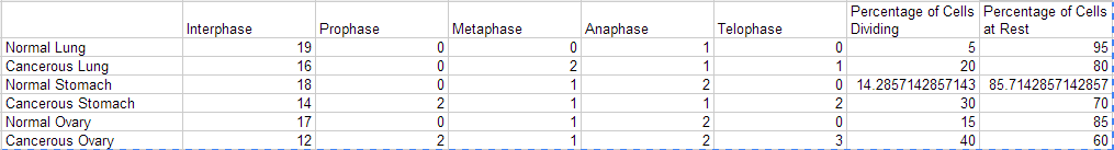
Below is a graph of the key data I collected:

VII. Journal Questions
- Cancer cells have "unique characteristics" not seen in normal cells including that cancer cells don't die, normal cells don't divide when in contact with other cells while cancer cells still divide, and cells don't perform the "intended' purpose.
- I believe that that the meiotic index is spent most in mitosis. Also, I believe that cancerous cells are in the higher range of the meiotic based on the notion that it spent little time in interphase and went directly to mitosis.
- I would have to say muscles have the highest meiotic index based on the fact that they can be "built up as they divide" and have the highest meiotic index due to the fact that they are always dividing.
VIII. Conclusion (Expected Results)
Based on the information I analyzed, a cancerous cell and normal cell could be told apart based on the amount of time spent in interphase. All the information taken concluded that the cancerous cells were in common in the way that they spent little time in interphase while the normal cells spent a longer period of time in interphase. Based off the information in my graph, the percentage of cells at rest is highest for normal cells at 95% while the lowest is 85%. In addition, the highest amount for cancerous tissues is 85% while the lowest the 15%. As you can see, this is the information I took based on the information of the normal and cancerous cells.
